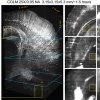
.jpg)
Neuroscience is still considered by many to be in its early stages of development, with researchers still trying to understand how events on the scale of neurons combine to produce high-level functionality that results in thinking, motion, etc. We reported last year on a new technique developed at Stanford University, called CLARITY, that turns brains into transparent objects that can be studied in 3D at various levels of resolution. Now the same team has further developed CLARITY to prevent damage to the preserved brains, retaining more of their detailed structures for study. Additionally, an imaging modality called light-sheet microscopy has been adapted to image the clarified brains more than 100 times faster than previously.
These advancements permit the imaging of a whole human brain at previously unprecedented resolution within about 220 days, as opposed to more than 80 years using the original CLARITY.
From the study abstract in Nature Protocols:
Here we describe protocols spanning multiple dimensions of the CLARITY workflow, ranging from simple, reliable and efficient lipid removal without electrophoretic instrumentation (passive CLARITY) to optimized objectives and integration with light-sheet optics (CLARITY-optimized light-sheet microscopy (COLM)) for accelerating data collection from clarified samples by several orders of magnitude while maintaining or increasing quality and resolution. The entire protocol takes from 7–28 d to complete for an adult mouse brain, including hydrogel embedding, full lipid removal, whole-brain antibody staining (which, if needed, accounts for 7–10 of the days), and whole-brain high-resolution imaging; timing within this window depends on the choice of lipid removal options, on the size of the tissue, and on the number and type of immunostaining rounds performed. This protocol has been successfully applied to the study of adult mouse, adult zebrafish and adult human brains, and it may find many other applications in the structural and molecular analysis of large assembled biological systems.
Source:Neuroscience
Clarity Method For Making Brains Transparent Now Much Faster, Allowing Visualization Of Even Smaller Details