GE Healthcare received FDA approval for its Invenia automated breast ultrasound system (ABUS), a novel device that can help physicians spot breast tumors when used in addition to mammography. Intended mostly for women with dense breast tissue, ABUS technology was successfully used to spot 35.7% more cancers than mammography on its own in a study used by the FDA to evaluate the device.
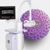
ABUS GE GE ABUS Automated 3D Ultrasound Breast Imaging System Approved in U.S.The automated scan takes about 15 minutes and operator-independent results are displayed as coronal view slices. Though attractive, the system doesn’t come supplied with the pictured giant flower.
Advanced algorithms automate the imaging process to help provide remarkable image quality and reproducibility from user to user, these include: Tissue Equalization, Nipple Shadow Compensation, Breast Border Detection and Chest Wall Detection. All of these are all designed to eliminate the distractions, and focus the radiologist’s attention on the most important data – the anatomy.
Operator workflow is smooth and easy with the Invenia ABUS high-resolution touchscreen display’s advanced Projective Capacitive Touch (PCT) technology. Its sleek, graphical user interface enhances the way you work. Tap and swipe colorful icons to quickly and easily maneuver through the Invenia ABUS exam.
The patented Reverse Curve? transducer offers extraordinary image performance, enhanced breast coverage and patient comfort. It is an extraordinary new class of transducer design conforms to a woman’s anatomy.
The scan head assembly includes Compression Assist, a feature which automatically applies up to 3 levels of compression to the breast for patient comfort, operator ease and image acquisition quality.
Images generated from the Invenia ABUS Scan Station are sent to the Invenia ABUS Workstation for interpretation, enabling fast, quick review and may be archived to PACS or external storage to optimize breast ultrasound workflow.
3D volumes are displayed in a patented, 2 mm thick coronal view slice from the skin to the chest wall using proprietary pattern recognition software. The result is a reading environment which allows for rapid and intuitive analysis of intricate breast anatomy and pathology.
source:GE Healthcare
GE ABUS Automated 3D Ultrasound Breast Imaging System