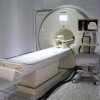
For MRI-guided breast biopsies, a scan is typically performed with the patient in the prone position and ultrasound is then used to guide the needle based on the scan while the patient is in supine position. This creates an imaging mismatch because, as everyone knows, the breast changes shape between the two positions, creating a lot of uncertainty when taking tissue samples.
Researchers at Germany’s Fraunhofer Institute for Biomedical Engineering and the Fraunhofer Institute for Medical Image Computing have developed a system called MARIUS (Magnetic Resonance Imaging Using Ultrasound) that solves this problem in an elegant fashion. Stick on ultrasound transducers are attached to the breasts, creating a reference map of the tissue. During MRI the scans are correlated to the ultrasound map and the patient is then moved to have the biopsy performed with the transducers still activated. So while the breasts may change shape, the map helps precisely locate suspicious tumors seen under MRI.
To realize this vision, Fraunhofer researchers are developing a range of new components. “We’re currently working on an ultrasound device that can be used within an MRI scanner,” says IBMT project manager Steffen Tretbar. “These scanners generate strong magnetic fields, and the ultrasound device must work reliably without affecting the MRI scan.” Ultrasound probes that can be attached to the body to provide 3D ultrasound imaging are also being developed by the team as part of the project.
The software developed for the technique is also completely new. “We’re developing a way to track movements in real time by means of ultrasound tracking,” explains MEVIS project manager Matthias Günther. “This recognizes distended structures in the ultrasound images and tracks their movement. We also need to collate a wide range of sensor data in real time.” Some of the sensors gather data about the position and orientation of the attached ultrasound probes while others track the position of the patient.
The team will showcase the entire concept and an initial demonstrator of the technology in November at the MEDICA 2013 trade fair in Düsseldorf at the joint Fraunhofer booth (Hall 10, Booth F05). The next version is set to be completed next year. Whereas the IBMT team is developing the hardware and new ultrasound techniques, the MEVIS working group is concentrating on the software.
source:Fraunhofer
New Ultrasound System Brings New Level of Precision to MRI-guided Biopsies

