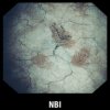
Olympus received clearance from the FDA to introduce its Narrow Band Imaging technology in the U.S. This is the only endoscopic light imaging system that helps resolve lesions without dyes or contrast agents by helping to see vessel networks, which are typically more dense in neoplastic tissues. This is done by illuminating the tissue with a combination of blue/green light and skipping the red component.
Specifically, the technology is indicated for assessing Non-Muscle-Invasive Bladder Cancer (NMIBC) in patients, but is not intended to supplant histopathological sampling for the definitive diagnosis.
Based on a weighted average, the aggregated FDA-reviewed studies show NBI has visualized NMIBC lesions in:
17 percent additional patients when compared with white light
24 percent additional tumors
28 percent additional carcinoma in situ (CIS or difficult-to-detect flat lesions)
Blue and green light are strongly absorbed by blood and appear darker than normal tissue. Blue light (415nm) highlights the shallow capillaries and green light (540nm) highlights deeper veins (type of blood vessel). NBI’s potential visualization of bladder cancer symptoms has been acknowledged by the medical community, but in 2013 a meta-analysis reviewed more than 30 disparate studies on the topic, enough to submit to the FDA.
NBI can be used for NMIBC in the office/clinic for cystoscopy (diagnosis) and in the O.R. or ambulatory surgical center for resection (tumor removal or Transurethral Resection of Bladder Tumor [TURBT]).
In-office, NBI provides new advantages to physicians because the most effective targeting technologies to-date could only be used in the O.R. and could only be used a single time per patient. With NBI, small tumors may be fulgurated in-office and prevent O.R. visits if visualized early enough, reducing costs associated with a costly-to-treat disease. In addition, NBI does not require the patient to take drugs or experience the discomfort of maintaining a full bladder compared with some treatments. NBI is available in both flexible and rigid endoscopes with no contra-indications. From the patient’s perspective, the equipment that uses NBI is the same as typical imaging equipment.
In the O.R., NBI can be used prior to resection to enable effective targeting of biopsies not seen under white light. During resection, NBI can be used to enhance visibility of tumor margins. By enhancing visibility of lesion boundaries, surgeons may be able to perform a more complete resection. Unlike technologies without FDA indications that strip out colors after taking the image with white light, NBI is filtered at the light source to allow application of only blue and green, for a deeper enhancement of vasculature.
Source:Olympus
Olympus Narrow Band Imaging for Cystoscopy