Nanolive, a spin-out company from Ecole Polytechnique Fédérale de Lausanne in Switzerland, has developed a revolutionary new 3D microscope. The 3D Cell Explorer microscope uses holographic and tomographic imaging technologies which allow scientists to quickly image cells in 3D without prior chemical preparation, saving significant time while producing some truly amazing nano-scale images.
At the heart of the microscope is a camera and lens mounted below the transparent sample container which is illuminated from above by a rotating 520nm beam. The microscope is capable of scanning at 200nm resolution in the xy plane and 500nm in the z-plane, with an 80μm field of view and a 30μm depth of field.
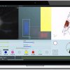
Once scanned, images and video of the cells are then reconstructed and processed by STEVE, Nanolive’s equally impressive software. STEVE is designed to use a standard graphics processor and allows the user to control the microscope, carry out interactive staining, perform quantitative analysis on the captured data, or export it for sharing or 3D printing. What is also highly impressive is the product design focus that has gone into both Nanolive’s hardware and software, which looks firmly set to disrupt the world of cell imaging and research.
The patented technology was developed over a number of years and details were published in 2013 in the journal Nature Photonics. STEVE, Nanolive’s software is available for download now and the 3D Cell Explorer microscope has been available for pre-order since last November. We know of at least one scientist who will be updating her lab wish list.
source:Nanolive
Holographic 3D Cell Explorer & STEVE Set to Disrupt Cell Imaging