Scientists at Purdue University are investigating a newly developed technology for monitoring the metabolism inside living cells, hopefully leading to a new diagnostic modality for spotting cancer and other diseases. The technique is an optimization of vibrational spectroscopic imaging by stimulated Raman scattering microscopy, souped up to be able to capture images at a high rate of speed.
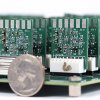
The technique relies on a device the scientists built, called a 32-channel tunded amplifier array (TAMP), that is able to image a tissue region every 32 µs. One interesting aspect of the new technology is that it is able to analyze large groups of cells as part of an entire organ, or just focus on individual cells passing through a flow cytometer.
“You can look at large numbers of cells from a patient’s blood sample to detect tumors, for example, and you can also look directly at organs with an endoscope,” said Cheng, Ji-Xin Cheng, a professor at Purdue University. “These capabilities will change how people use Raman spectroscopy for medicine. There are many organelles in each cell, and spectroscopy can tell us what’s in the organelles, which is information not available by other techniques.”
Incorporated with multivariate curve resolution analysis, our platform allows compositional mapping of lipid droplets in single live cells, observation of intracellular retinoid metabolism, discrimination of fat droplets from protein-rich organelles in Caenorhabditis elegans, spectral detection of fast flowing tumor cells and monitoring drug diffusion through skin tissue in vivo. The reported technique opens new opportunities for compositional analysis of cellular compartment in a microscope setting and high-throughput spectral profiling of single cells in a flow cytometer setting.
source:Purdue University
Vibrational Spectroscopic Imaging