

An imaging revelation
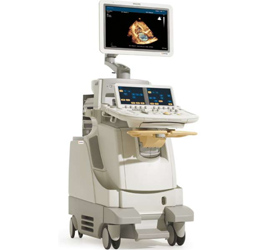
Combining unprecedented 2D and 3D image quality in the same transducer and a host of easy-to-use quantification, clinical performance, and information management tools, the new iE33 xMATRIX echo system addresses the clinical needs of managing patients with cardiac disease, including heart failure, valvular disease, and congenital heart disease.
The iE33 xMATRIX reveals:
Versatile X5-1 transducer
With 3,000 elements and breakthrough PureWave xMATRIX technology, the X5-1 supports virtually any cardiac ultrasound exam, including 3D, 2D, color flow, M-mode, PW/CW Doppler, Tissue Doppler imaging, and contrast-enhanced exams. It delivers outstanding image quality and a new handle design with relaxed grip to reduce user fatigue and improve scanning stability.
Use iRotate to electronically rotate the image to get the best view possible, and more easily obtain challenging views, such as apical two-chamber, while reducing foreshortening.
Reduce your stress in stress echo
The new Philips iE33 xMATRIX automates stress echo exams, so they’re fast and consistent. Use iRotate Stress Echo in combination with the X5-1 transducer to complete an entire stress echo protocol. You can include acquisition of 2-chamber, 3-chamber, and 4-chamber 2D images all from the standard windows following peak patient exertion—without rotating the transducer.
As you move electronically around the heart capturing baseline images, making any necessary small angle adjustments, these adjustments will be saved, along with gain and depth settings, for instant recall when you are in your post-exercise or -pharmacologic stage. A much less stressful stress echo solution that benefits both you and your patients.
With CMQ-Stress, you can quantify 2D stress echo studies and communicate global and regional LV function during each stage. And Live 3D stress echo with iSlice lets you add volume acquisition to your stress protocol and slice it later for the best views and content to make informed diagnoses—with confidence.
Accurate 2D and 3D LV function and regional wall motion assessment
The QLAB CMQ plug-in enables assessment of left ventricle global function and regional wall motion, deformation and timing. The improved speckle-tracking algorithm provides you with objective quantification of global and regional wall abnormalities.
Rapidly acquire more clinical data from 3D exams with Cardiac 3D Quantification Advanced (3DQ Advanced). Accurate, semi-automated analysis of true LV volumes, and with the iE33 xMATRIX system can provide routinely accurate ejection fraction in under 1 minute.
More information for interventional procedures
Live 3D TEE (transesophageal echo) is providing clinical cardiologists, cardiac surgeons, anesthesiologists, interventional cardiologists, and echocardiographers with views of cardiac structure and function seen for the first time. Capturing a 3D volume is quick, accurate, reproducible, and quantifiable. You can view the 3D heart in real time as well as review the 3D volume at any time, select any plane for interrogation, and perform extensive quantification. It’s more information for diagnosis and treatment planning.