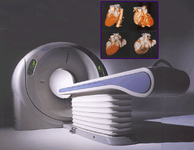

The Aquilion is a multislice Helical CT system that supports whole-body scanning. The system generates 128 slices per second using the Selectable Slice-thickness Multi-row Detector (SSMD). In addition, the high-speed rotation mechanism and the fast reconstruction unit of the system allow quick image acquisition to further improve throughout in CT examinations.

Special features:
Wide range of slice thicknesses
This detector is provided with detector elements that have high-power and uniform output characteristics, permitting scanning with a minimum scan slice thickness of 0.5 mm. Image slice thicknesses of up to 10 mm can be selected based on the objectives of the examination.
Advanced image reconstruction
This system employs the helical reconstruction technique TCOT (True cone-beam tomography), which precisely compensates for the cone angle based on the Feldkamp method, thus minimizing artifacts related to the cone angle and providing accurate, high-resolution isotropic images over the entire imaging field. In addition, MUSCOT (multislice cone-beam tomography) reconstruction, which has received accolades in 4-row multislice CT, can also be used.
High-quality images
The system achieves low-contrast detectability of 2 mm at 0.3% and high-contrast resolution of 0.35 mm. Multislice helical CT can be performed using thin slices in routine examinations so that high-precision 3D and MPR images can be generated from the high-resolution voxel data. In CT angiography of the brain, for example, a 40-mm distance can be scanned in about 3 seconds at a slice thickness of 0.5 mm.
High patient throughput
In combination with fast scanning, the high-power 60-kW X-ray generator and 7.5 MHU X-ray tube permit data for routine examinations, biopsy procedures, and 3D image generation to be acquired in one scan. Images of various slice thicknesses can be generated. For example, it is possible to reconstruct 5 mm to 10 mm slice images for routine examinations, 2 mm to 5 mm slice images for detailed examinations and 0.5 mm or 1 mm slice images for 3D image generation from the data acquired in a 0.5 mm or 1 mm slice helical scan.
Tilt Helical scanning
Helical scanning with gantry tilt, from 30° forward to 30° backward, is available because the system employs the TCOT reconstruction technique.
SureFluoro (option)
Conventional CT fluoroscopy shows only a single slice, but SureFluoro (Multislice CT fluoroscopy) permits real-time image reconstruction to display 3 images obtained by combining data from the SSMD. SureFluoro significantly improves operability in biopsy and interventional procedures.
SureExposure
It is possible to perform helical scanning while continuously varying the X-ray tube current in order to optimally control the X-ray dose for the region being scanned. This makes it possible not only to reduce the patient exposure dose, but also to obtain uniform image quality in the cross-sectional images obtained. Furthermore, there is also an exposure reduction mode that reduces the dose to the patient by about 40%, in addition to the conventional mode. This allows controlling the exposure dose according to the purpose of the examination.
Dual monitor system
To provide a comfortable examination environment, this system employs a dual monitor system. A scan system monitor and an image processing system monitor can be operated independently. This permits parallel processing, improving the examination efficiency.
?
True multi-tasking capability
The image reconstruction system is completely separate from the scan system, enabling continuous image reconstruction even during scanning. This makes it possible to increase the workflow speed dramatically.
Improvements in volume data workflow
This system includes a function for automatically generating MPR images after completion of scanning. A high-speed image feeding function is also provided. Image display, image switching, filming, and archiving can be performed for the generated MPR images using the same user interface as for axial images. In addition, the acquired voxel data can be processed as a single voxel data set (rather than as a large number of axial data sets), permitting storage, loading, and list display to be performed in units of volume data sets. These functions are useful for image analysis. The generated MPR images can be automatically transferred to multiple destinations such as an imager (slice) switching can be performed using the hard keys on the keyboard, which is very useful for image analysis.