
MR-Acquisition and Processing in Preclinical Research and Material Science

Main Features
- Intuitive routine workflow
- Application-oriented, ready-to-use protocols
- Self-acting, method-specific scanner adjustments
- Automatic instrumentation recognition
- Parallel acquisition option for all suitable acquisition techniques with automatic generation of composed images/spectra
- Half-Fourier (Partial-Fourier) encoding
- Self-gated cardiac MRI using IntraGate™ enables continous scanning under steadystate conditions
- Real-time display of acquired and reconstructed data
- Sophisticated data archiving including DICOM export
- Development environment with powerful tools for rapid prototyping of user-defined experiments and professional method implementation
Reconstruction Features
- Push-button GRAPPA reconstruction for all parallel imaging methods
- Sum of squares and phase-sensitive, phased-array reconstruction strategies
- Phased-array spectroscopy reconstruction
- Partial FOURIER reconstruction
- EPI reconstruction with efficient ghost suppression
- Navigator techniques to reduce motion artifacts in EPI and SPIRAL
- DTI evaluation
Processing Features
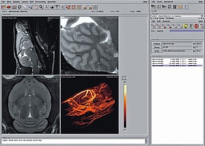
A rich palette of image analysis and visualization tools allow the user to extract more complex information from 2D or 3D images. Besides basic functionality like magnification, ROI analysis and geometrical measurements, the following features have proven highly invaluable:
- 2D and 3D region growing, e.g. for image segmentation
- Export of histogram values
- Display and analysis of time-course´data with the fitting tool “ISA”
- Frame-selective loading of image sequences for display, e.g. either timecourse frames or slices for a multi-slice movie dataset
- 3D visualization with surface rendering Image mask inversion
What's New with ParaVision 5.1
Three-dimensional UTE
Three-dimensional ultra-short TE imaging. Like the two-dimensional UTE introduced with PV5.0, UTE3D acquires the k-space radially during the rising ramp of the readout gradient. The 3-dimensional sampling eliminates the need of slice selection and allows a significant reduction of the echo time. It is possible to achieve TE values as short as 10 microseconds. The reconstruction is based on the k-space gridding with automatically measured trajectories and produces artefact-free images without manual trimming of the sequence.
Zero-TE imaging
Three-dimensional radial sampling using a hard RF pulse applied during the readout gradient. Due to the absence of a delay between the excitation and the k-space centre, the effective "echo time" of the sequence is zero. ZTE can be used for materials of extremely short T2* and in the presence of high susceptibility gradients. The acquisition dead time occurring in this kind of techniques is handled by a special algebraic reconstruction step.
T1 EPI
A variant of Echo-Planar Imaging in which a train of images is acquired in a single inversion-recovery experiment allowing a snapshot acquisition of T1 maps using the principle of the Look-Locker sequence. The method uses k-space segmentation and cyclic slice permutations to achieve required time resolution in multi-slice experiments.
T2* EPI
A multi-echo variant of EPI providing a train of images with different radient echo times in a single shot. The method is useful for fast T2* mapping and for quantitative fMRI experiments.
T2 EPI
A multi-echo variant of EPI based on the CPMG sequence for fast T2 mapping.
FAIR RARE
A pulsed arterial spin marking method for perfusion imaging based on the FAIR preparation and a fast RARE readout. The advantage of this method compared to the existing FAIR_EPI is the lack of distortions and signal voids caused by ma