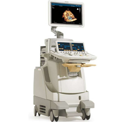

iE33 xMATRIX systemoffers expanded clinical applications in cardiac ultrasound imaging

Royal Philips Electronics announced enhancements to the iE33 xMATRIX cardiac ultrasound system designed to provide a more complete imaging solution for adult echocardiograms. In addition to the ergonomic design of the new X5-1 transducer, which aims to improve comfort and efficiency for the clinician, the system has been developed to help advance patient care by enabling expanded cardiac-related diagnostic capabilities related to ischemic disease detection, structural heart disease assessment, as well as systolic and diastolic heart failure and arrhythmia. These new features, which benefit both clinicians and patients, will be displayed at the European Society of Cardiology (ESC) Congress in Stockholm, August 28 through September 1.
“The new iE33 xMATRIX ultrasound system represents a significant breakthrough in ultrasound; technology that gives clinicians new diagnostic tools and helps them examine patients more quickly,” said Andrew Hatt, interim general manager, Ultrasound, for Philips Healthcare. “In addition, clinicians who have experienced scanning fatigue will appreciate that the X5-1 transducer combines the ergonomics of 2D transducers with the three-dimensional technology they need, to make the most informed patient care decisions possible.”
Continuing the tradition of simplified, patient-focused healthcare solutions, new features of the iE33 xMATRIX ultrasound system include outstanding 2D and 3D image quality utilizing a single transducer, near instantaneous acquisition of 3D volumes, new 3D workflow tools and visualized color Doppler flow pattern during 3D exams. With the X5-1 and iRotate, a clinician can more easily obtain challenging 2D views, such as apical two-chamber. Rather than manually rotating the transducer and searching for a window that isn’t obscured by ribs, rotation is achieved electronically to maintain the best acoustical window. When used in stress echo, iRotate allows clinicians to complete an entire stress echo protocol from the standard windows following peak exertion without rotating the X5-1 transducer.
The iE33 xMATRIX ultrasound system also offers a 3D stress echo solution that can be incorporated with conventional 2D stress echo. After acquiring a volume, clinicians can then use the system’s advanced iSlice software to obtain short axis views of the heart’s apex. This constitutes additional clinical information that is not routinely obtained by ultrasound.